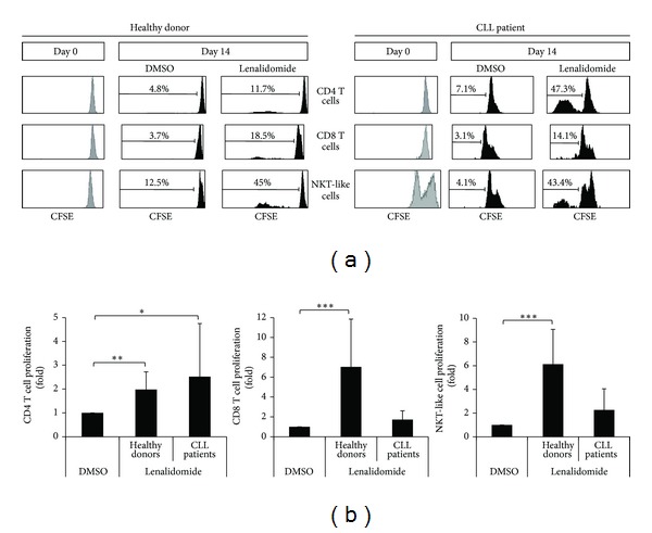
Figure 4.

Effect of lenalidomide on the proliferation of T cell subsets. (a) The histograms show the CFSE expression of CD4 T cells, CD8 T cells, and NKT-like cells before and after stimulation with lenalidomide. PBMCs were labeled with CFSE and cultured with 1 μM lenalidomide or DMSO for 14 days. CFSE expression in CD4 T cells (CD3+CD4+), CD8 T cells (CD3+CD8+), and NKT-like cells (CD3+CD8+CD56+) was examined by flow cytometry. One representative CLL patient and one donor are shown. (b) The figure shows the compilation of the results obtained from CLL patients (n = 17) and donors (n = 10). Results are expressed as the fold induction of the percentage of proliferative CD4 T cells, CD8 T cells, and NKT-like cells of lenalidomide-treated cells relative to the vehicle-treated control. (*P < 0.05; **P < 0.01; ***P < 0.001, Mann-Whitney U test).
