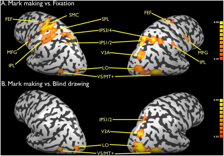
Figure 2. Brain activations for mark making.
a) Mark making vs. fixation. b) Mark making vs. blind drawing. Data are corrected for multiple comparisons using FDR p<0.01. Activations shown in Figures 2–5 are rendered onto an inflated brain of one of the subjects in the study (Subject 4) as normalized into Talairach space. The color bars in Figures 2–5 reflect the t score of the activated voxels for a given contrast. Abbreviations: FEF: frontal eye fields; IPS1/2: segments 1 and 2 of the intraparietal sulcus; IPS3/4: segments 3 and 4 of the intraparietal sulcus; MFG: middle frontal gyrus; MT: middle temporal; SMC: sensorimotor cortex; SPL: superior parietal lobule.

