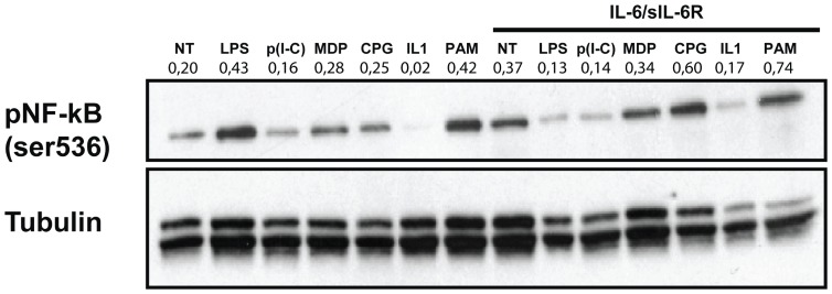
Figure 4. Increased p65 NF-κB activation in response to inflammatory stimuli in human PBMCs pre-exposed to IL-6/IL-6R.
Western blots showing the expression of phospho–p65 NF-kB (Ser536) in total lysates from human PBMCs pre-exposed to IL-6 (10 ng/ml) in combination with sIL-6R (125 ng/ml) for 1 hour and stimulated with LPS, poly(I-C), MDP, CpG, IL-1β, PAM (used at the same concentrations as in Figure 2) for 90 minutes. Expression of α-tubulin was used as a loading control. Results of densitometric analysis are shown above each blot. Data are representative of 3 independent experiments.

