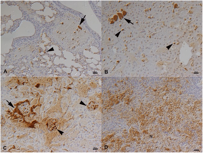
Figure 1. Naturally F. tularensis sp. holartica infected field vole that had been trapped and euthanized [27].
A. Lung with bacterial clumps in vessel lumina (arrow) and bacterial aggregates in capillaries (arrowhead). B. Liver with bacterial clumps in sinusoids (arrow) and smaller aggregates within Kupffer cells (arrowheads). C. Kidney with bacterial aggregates in larger vessels (arrow) and glomerular capillaries (arrowheads). D. Spleen with abundant bacterial clumps, in association with necrosis (N), in the red pulp. Horseradish peroxidase method, Papanicolaou’s hematoxylin counterstain. Bars = 20 µm.

