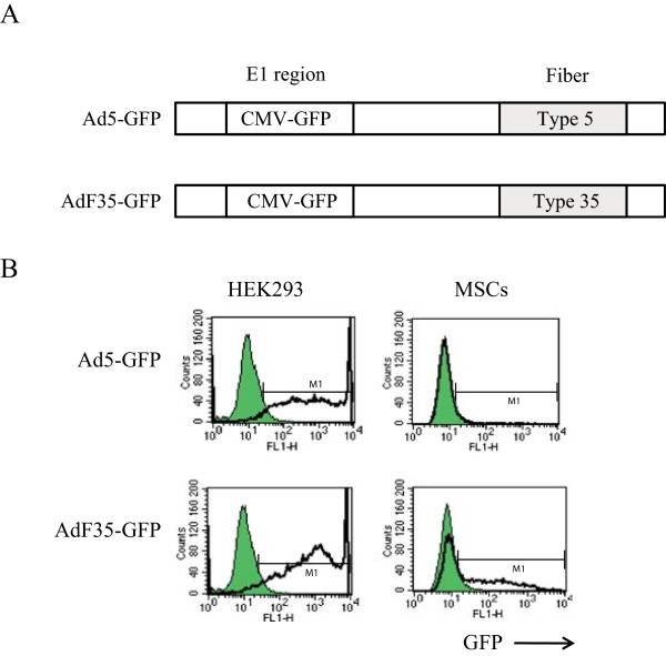
Figure 3.

Infectivity of Ad to MSCs. (A) Schematic structures of Ad5-GFP and AdF35-GFP. The E1 region was replaced with the CMV promoter-linked GFP gene. (B) Representative flow cytometry profiles of HEK293 cells and MSCs that were infected with Ad5-GFP or AdF35-GFP. M1 indicates positively stained population, and shaded areas and bold lines show uninfected and infected cells, respectively.
