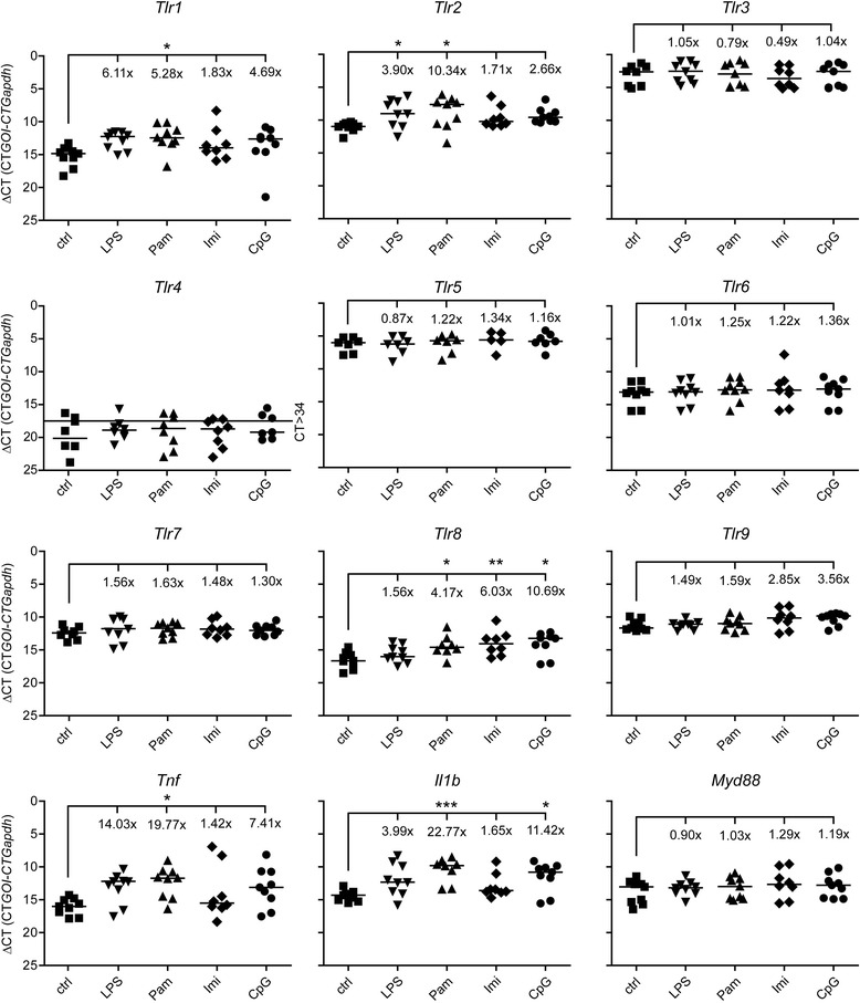
Figure 6.

mRNA expression of TLRs in response to activation of a single TLR in the CNS . C57BL/6J mice were injected intrathecally with 10 μg LPS (n = 9), 10 μg Pam3CysSK4 (Pam, n = 9), 10 μg imiquimod (Imi, n = 8), or 10 μg CpG ODN (CpG, n = 9). Intrathecal injection of 0.9% NaCl (ctrl, n = 9) served as control. After 12 h, brain tissue was analyzed for mRNA expression of TLRs and proinflammatory molecules, as indicated, by quantitative real-time PCR. Data are presented as delta CT (dCT = CTgene of interest – CTGapdh) of each mouse with median per group on reverse scale to visualize changes in mRNA levels. Fold increase (2-ddCT, with fold change >2 and <0.5 expressing biological significance) was calculated with the median of each group, setting the control to 1; ANOVA of log2 transformed dCT values followed by Bonferroni post-hoc test of control vs. treatment. P* <0.05; P** <0.005; P*** <0.001.
