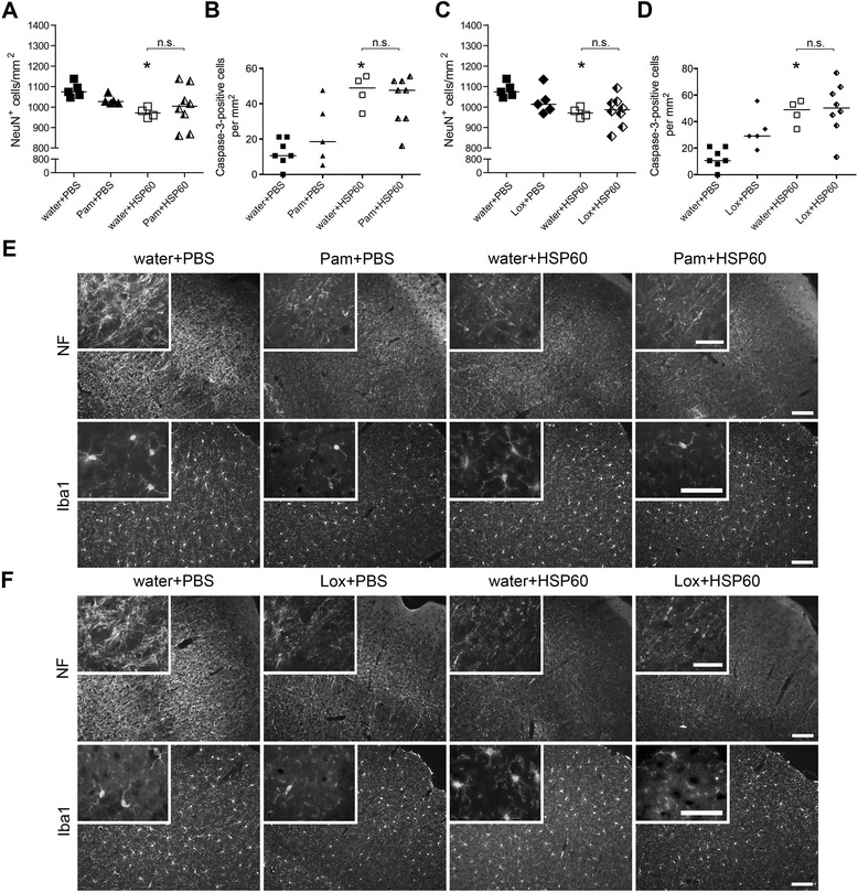
Figure 8.

Subsequent challenge with HSP60 does not exacerbate CNS damage induced by exogenous TLR ligands. C57BL/6J mice received two subsequent intrathecal injections. On day one 40 μL water (carrier), 40 μg Pam3CysSK4 (Pam), or 136 μg loxoribine (Lox) were injected. After 24 h, 40 μL PBS or 40 μg HSP60 were injected additionally (water + PBS n = 7; water + HSP60 n = 4; Pam + PBS n = 5; Lox + PBS n = 5; Pam + HSP60 n = 8; Lox + HSP60 n = 8). After a further 3 days, brains were analyzed by immunohistochemistry. (A, C) NeuN+ cells of the cerebral cortex were quantified, and the mean is expressed as NeuN+ cells per mm2; median with Kruskal-Wallis test followed by Bonferroni post-hoc test of control (water + PBS) vs. treatment or between the respective treatments, as indicated. P* <0.05; n.s.: not significant. (B, D) Brain sections of injected mice were immunostained with an Ab against activated caspase-3. Cortical cells positive for activated caspase-3 were quantified. The mean of six high power fields per cerebral cortex is expressed as caspase-3+ cells per mm2; median with Kruskal-Wallis test followed by Bonferroni post-hoc test of control (water + PBS) vs. treatment or between the respective treatments, as indicated. P* <0.05; n.s.: not significant. (E, F) Representative micrographs of the cerebral cortex immunostained with neurofilament Ab (NF) and Iba1 Ab to mark axons and microglia, respectively. Scale bar, 100 μm, inset 50 μm.
