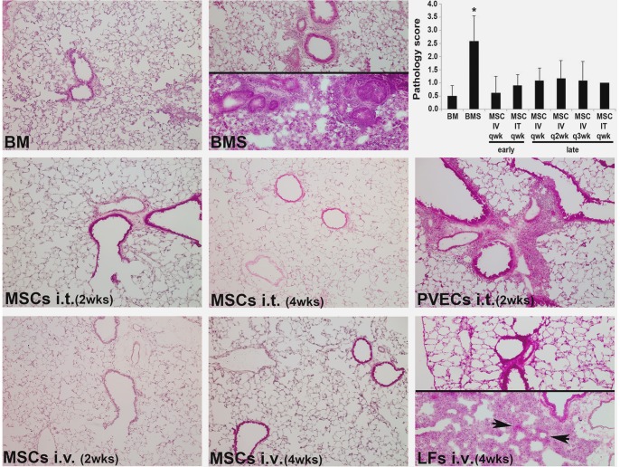
Figure 3. Administration of MSC post-BMT reduces OB histopathology.
Representative H&E stained lung cryosections of day 90 post-BMT shown for mice receiving T cell-depleted bone marrow only (BM); BM and allogeneic splenocytes (BMS; OB group). The split panels show the range of injury in this model. MSCs IV or IT as indicated, beginning at 2 or 4 weeks and given weekly; PVECs IT beginning at 2 weeks and given weekly; LFs IV beginning at 4 weeks and given weekly until day 90. The split panels show the range of injury seen in this group similar to the BMS group. Original magnification 200× (20× objective lens). The top right corner panel shows semiquantitative pathology scores; n = 4–6/group pooled from 4 experiments.

