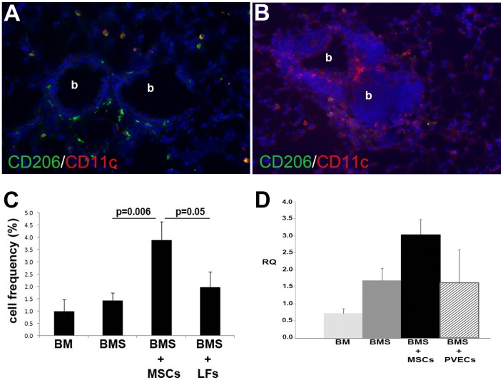
Figure 5. Increased expression of markers of AAMs in lungs of mice given MSC post-BMT.
Starting at 4 weeks post-BMT, MSCs (A) or LFs (B) were administered IV weekly and evaluated on day 90 post-BMT. Lung cryosections were immunofluorescently stained for CD11c with CD206 (A, B) and frequency of CD206+ cells was determined as a percent of total nucleated cells (data in C shown as mean ± SE, n = 5–7/group pooled from 2 experiments). Images are at 400X total magnification; “b” indicates bronchiolar airway. In D, cell therapy (IT, weekly) was started at 2 weeks and lungs examined on day 90 post-BMT by qRT-PCR for CD206 (n = 3/group).

