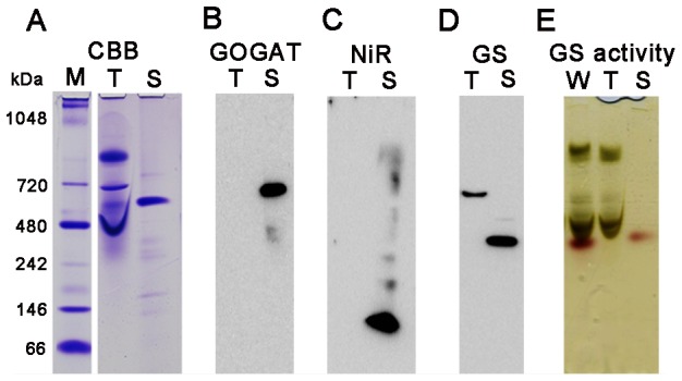
Figure 5. Protein complexes in spinach chloroplasts analyzed by using Native PAGE.

CBB staining (A), western blots (B–D) and in-gel GS activity staining (E) after Native PAGE analysis of thylakoid (T) and stroma (S) proteins extracted from spinach chloroplasts. For in-gel GS activity assay, proteins extracted from whole chloroplasts (W) were also used. Lane M stands for the protein standard markers, but the migration distance does not necessarily correlate with the molecular mass in this Native PAGE. Samples loaded were derived from 10 µg (A–D) and 20 µg (E) on a chlorophyll basis of chloroplasts whose proteins were extracted.
