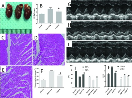
Figure 3. Effects of shIGF1R administration on cardiac hypertrophic process in vivo.

(A) Morphological characteristics of isolated heart from different groups. (B) Ratio of HW/BW in different group,*P<0.05. Histological evaluation the cross-sectional area of heart tissue from normal group (C), shIGF1R administrated group (D), saline-treated group (E), Magnification=200×. (F) shIGF1R administration attenuated the cross-sectional area compared with control, *P<0.05. Echocardiographic evaluation of heart function in normal mice (G), hypertrophy (H), shIGF1R-treated mice (I). (J) shIGF1R administration increased the fractional shortening of left ventricle (FS%) and EF%,*P<0.05 versus control. (K) Parameters of cardiac hypertrophy:LVDd;d, diastolic left ventricle internal dimension; LVDd;s were evaluated, *P<0.05 versus control.
