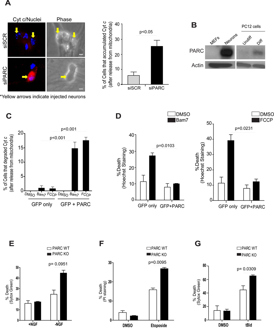
Figure 5.
(A) One day after micro-injection with siSCR or siPARC, sympathetic neurons were deprived of NGF for 24 hours in the presence of QVD-fmk (25 µM) and status of cyt c was determined by immunofluorescence and quantified. Error bars represent ± SD for triplicate experiments. (B) Western blot analysis comparing the levels of PARC between MEFs and sympathetic neurons as well as between undifferentiated (Undiff) and differentiated (Diff) PC12 cells. (C, D) HeLa cells were transfected with pCDNA3 empty vector or pCDNA3-HA-PARC and pEGFP. Three days later cells were treated with Bam7 or FCCP for 10 hours and the status of cyt c was determined by immunostaining (C) and cell death was quantified by nuclear morphology using Hoechst staining (D). (E) PARC knockout and wild-type neurons were deprived of NGF for 10 hours or treated with 10 µM Etoposide for 96 hours (F). Death was measured by the vital dyes Sytox Green or Propidium Iodide. Error bars represent ± SD for triplicate experiments. (G) PARC knockout and wild-type neurons were deprived of NGF in the presence of cycloheximide for 48 hours and then microinjected with tBid (8 mM). Death was measured by Sytox Green. Error bars represent ± SD for triplicate experiments.

