
Figure 2.
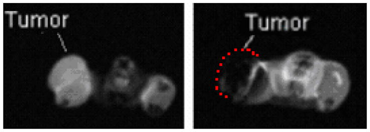
Left: The initial MRI scan of mouse 1 clearly distinguishes the tumor from the rest of the flank. Right: The scan taken after a direct injection of IONP with 7.5 mg Fe/gram of tumor shows significant quenching of the signal. Tumor location in image at right is indicated by dashed line. Imaging parameters: spin-echo, TR = 1000 ms, TE = 14.52 ms FOV = 60 × 60 mm, acquisition matrix = 128 × 128, 10 slices, slice thickness = 2mm, one signal average.