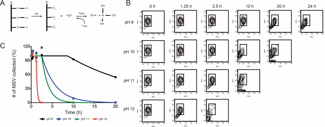
Figure 5. Degradation effect of MSV in varying pH environments.
A) Schematic showing possible interactions between the MSV surface and hydroxyl groups. Hydroxyl ions of an alkaline solution weaken the surface of the MSV forming bioinert silicic acid. B) Flow cytometry exhibiting distinct patterns and changes in size (forward scatter) and shape (side scatter) exposed to the varying pH environments. C) Flow cytometer counts collected at various time points to show degradation rates of MSV at pHs between 9 and 12.

