
Figure 1. Casp6 activation in axons is selective to NGF deprivation.
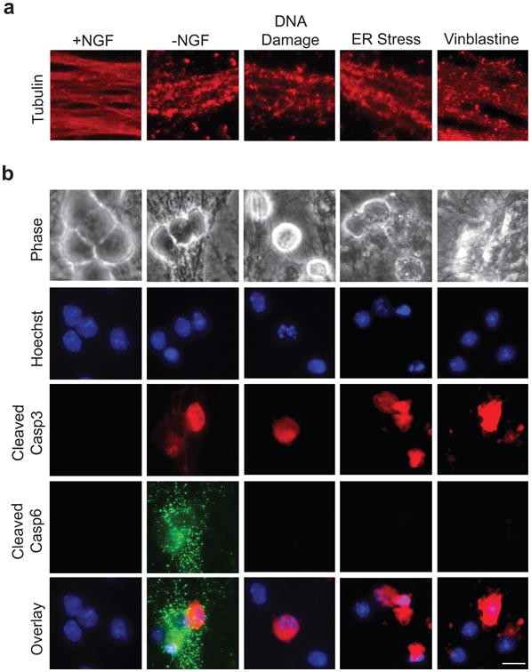
(a) Wild type (WT) sympathetic neurons (5 DIV) were treated with the following conditions to induce axon degeneration, respectively: NGF deprivation (-NGF), 20 μM etoposide (DNA Damage), 2.5 μM tunicamycin (ER Stress), and 1 μM vinblastine (to induce microtubule destabilization). NGF-maintained neurons served as controls. Neurons were fixed and immunostained for tubulin upon axon beading and fragmentation. (b) Neurons were treated as described in (a) and immunostained for cleaved Casp3 and cleaved Casp6. Nuclei were labeled with Hoechst. Scale bar, 20 μm.