
Figure 3. Axon-selective degeneration is Apaf-1-independent but requires Casp9 and Casp3.
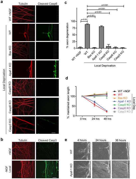
(a) Sympathetic neurons (5 DIV) from mice deficient for Bax, Apaf-1, Casp9, Casp6, and Casp3 were locally deprived in microfluidic chambers. NGF-maintained and locally deprived WT littermate neurons served as controls. Neurons were immunostained for tubulin and cleaved Casp6. (b) WT neurons were locally deprived for 24 hours and probed for cleaved Casp3. NGF-maintained neurons served as controls. (c) Quantification of axon degeneration for conditions shown in (a). (d) Quantification of maintained axon length over time during local deprivation for all conditions shown in (a). All data represent the mean ± s.e.m. (n=3). (e) Phase images of the same axons over a 36-hour timecourse of local deprivation demonstrate similar degeneration kinetics between WT and Apaf-1-deficient axons. All p values were calculated using an unpaired t-test. All Scale bars, 50 μm.