
Figure 4. Local deprivation triggers cytc release in axons.
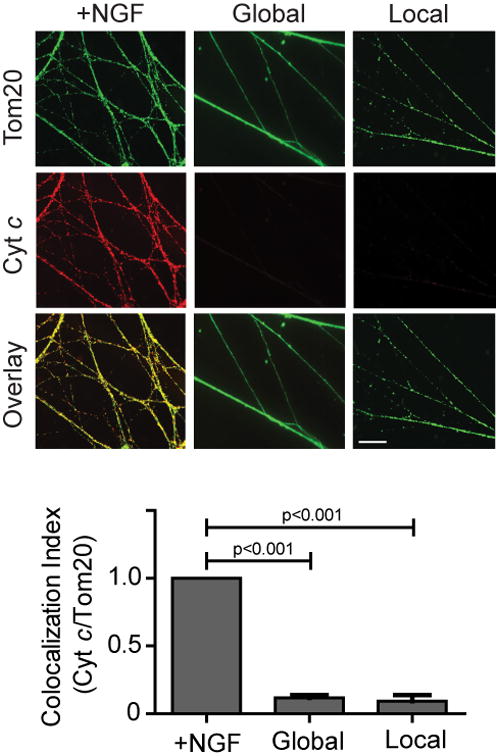
Neurons (5 DIV) were NGF-maintained, globally deprived, or locally deprived in the presence of pan-caspase inhibitor (25 μM QVD) for 36 hours and probed for cytc and the mitochondrial marker Tom20. Quantification of the colocalization index (ImageJ) for cytc and Tom20 is shown below. Data represent the mean ± s.e.m. (n=3). Scale bar, 10 μm. All p values were calculated using an unpaired t-test.