
Figure 3.
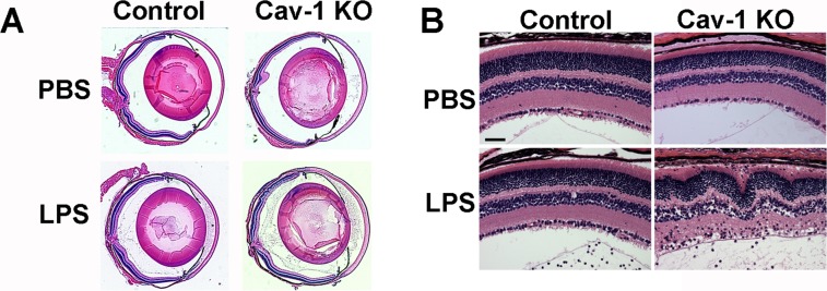
Histologic analysis of LPS-induced inflammation in Cav-1 deficient eyes. (A) Representative sections of whole globes (A) and regions of retinas (B) in LPS- and PBS-injected Cav-1 deficient and control mouse eyes. Similar anterior segment and vitreous infiltrates were observed but regions of Cav-1 KO retinas displayed more inflammatory disruption than controls. Scale bar: 50 μm.