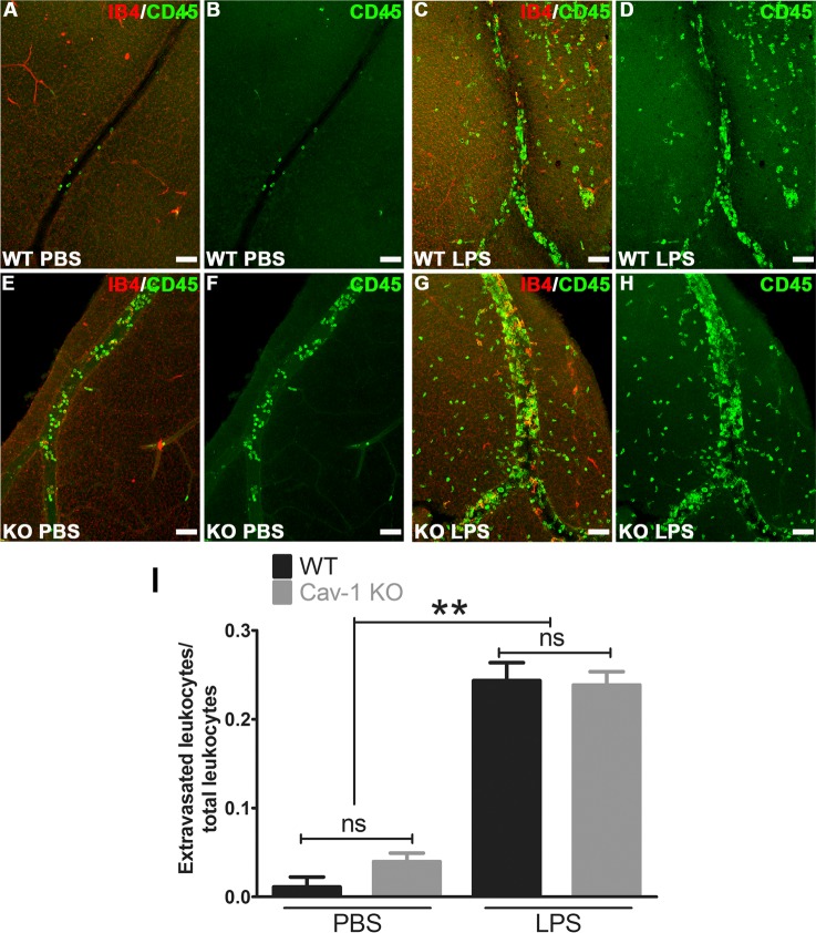
Figure 5.
Ratio of extravascular/total CD45-positive immune cells was not different between LPS-challenged Cav-1 KO and WT retinas. Retinal flatmounts were stained with CD45 (green) and isolectin-B4 (IB4, red). (A–H) Representative confocal stacks of whole mounts from WT and Cav-1 KO retinas focused on retinal veins in the superficial layer. (I) Results of quantification of ratios of extravascular/total immune cells (n = 3, mean ± SEM, **P < 0.01, *P < 0.05, ANOVA with Newman-Keuls post hoc test). Scale bar: 50 μm. ns = not significant.

