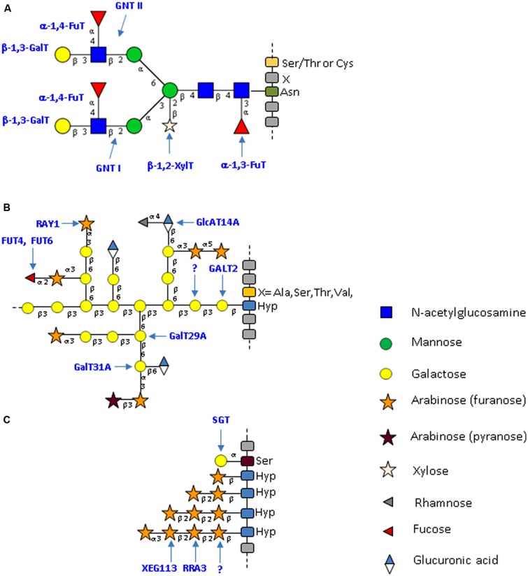
FIGURE 1.
Typical structure of plant N- and O-glycans from cell wall proteins. (A) Specific complex-type N-glycans attached to plant glycoproteins. This N-glycan results from the action of a plant-specific repertoire of glycosyltransferases that lead to the formation of a glycan bearing plant-specific glyco-epitopes such as a core β-1,2-xylose; a core α-1,3-fucose and a Lewisa antennae (Lerouge et al., 1998; Wilson et al., 2001b; Bardor et al., 2003, 2011). The N-glycan structures presented here are drawn according to the symbolic nomenclature adopted by the Consortium for Functional Glycomics (Varki et al., 2009). (B) Schematic representation of O-glycans (type II arabinogalactan) attached to AGPs. These glycans predominantly consist of arabinose and galactose. Minor sugars, such as glucuronic acid, fucose or rhamnose, are also present. The O-glycans are attached to non-contiguous Hyp residues. The model presented is modified from Tan et al. (2010) and Tryfona et al. (2012). (C) Schematic representation of O-glycans attached to plant EXT. These glycans consist of short chains of arabinose and on single galactose residues. The O-glycans are attached to contiguous Hyp residues. The model presented is modified from Saito et al. (2014). Yellow circle: galactose; green circle: mannose; blue square: N-acetylglucosamine; star: white xylose and red triangle: fucose; gray triangle: rhamnose; orange star: arabinose (furanose); purple star: arabinose (pyranose); blue/white diamond: glucuronic acid.

