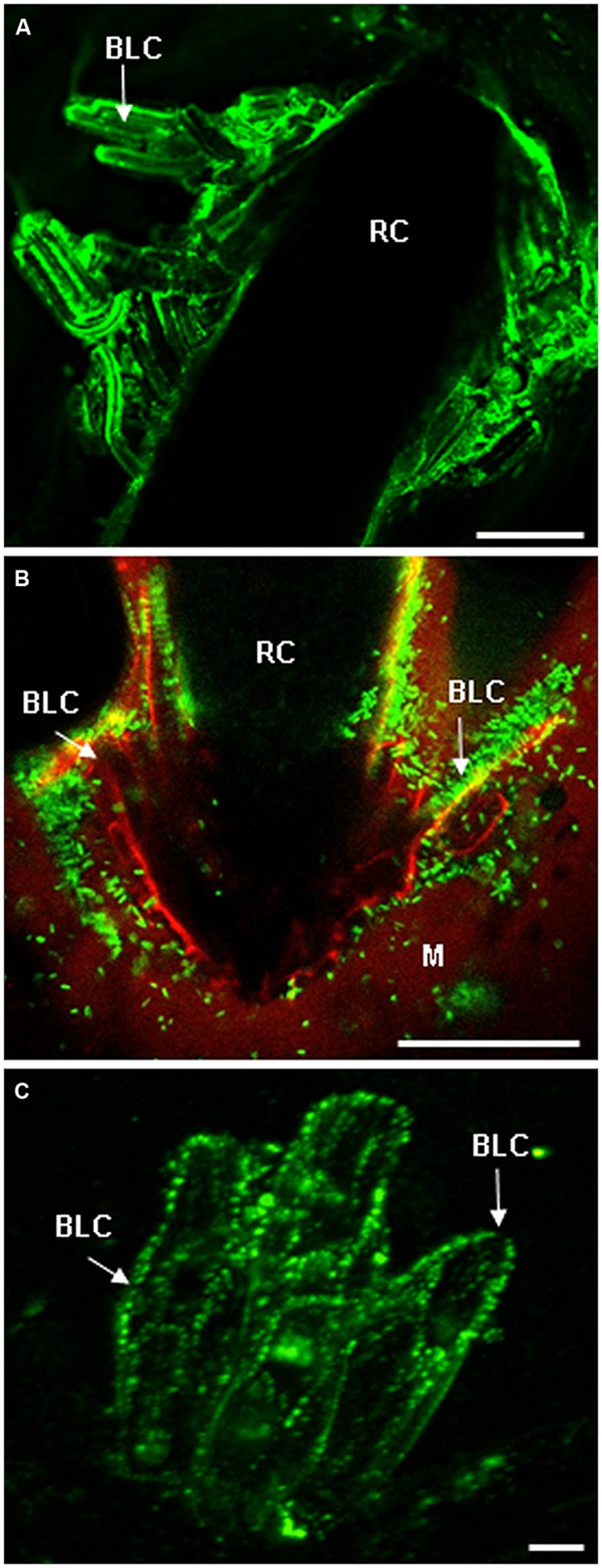
FIGURE 2.
Root cap (RC) and border cells are both enriched in AGP and EXT epitopes. (A) Immunostaining of AGP epitopes at the surface of RC and border-like cells of Brassica napus with the mAb JIM8 (from Cannesan et al., 2012 with permission). Root border-like cells are produced and released from the RC. (B) Micrographs showing the association between root border-like cells from Arabidopsis thaliana and Rhizobium sp. YAS34-GFP. The GFP-expressing bacteria appear green at the root surface (from Vicré et al., 2005 with permission). This association is AGP-dependant as demonstrated in Vicré et al. (2005). (C) Fluorescent micrographs of root border-like cells from flax (Linum usitatissimum) immunostained with the monoclonal antibody LM1 specific for EXT epitopes (from Plancot et al., 2013 with permission). Bars = 20 μm (A), 50 μm (B), and 8 μm (C). BLCs, border-like cells; M, mucilage; RC, root cap.

