Figure 3.
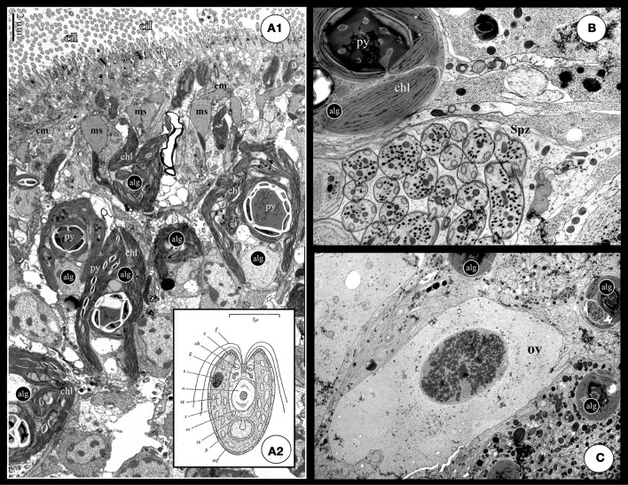
(A1) TEM cross section of a S. roscoffensis adult. On the top of the image, the ciliated (cilia: cil) epidermis is visible above a net of both circular and longitudinal muscles (cm and lm). Algal cells (alg) are in contact with the acoel cells below the muscles layer. Algal cells exhibit two specific features, the chloroplasts (Chl) and the pyrenoid with starch accumulation on the outside (the white mass). Note some algal extensions close to the epidermis, above the muscle boundary. (A2) Detailed scheme of the free-living unicellular green alga, Tetraselmis convolutae (modified from Parke and Manton, 1967): c, chloroplast; f, flagellum; g, golgi body; m, mitochondrion; mf, muciferous body; n, nucleus; p, pyrenoid; rb, refractive body of unknown nature; ss, starch shell; s, stigma; st, stroma starch; t, theca. (B,C) These two pictures exhibit a clear physical proximity of germinal cells, respectively sperm (Spz in B) and ovocyte (ov in C), and algae (alg). Such a situation could increase possible horizontal gene transfers.

