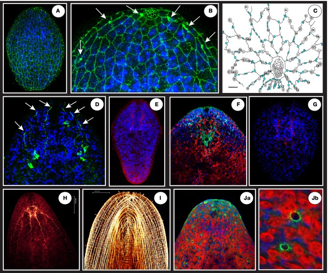
Figure 4.
(A) A net-like structure labeled by a VDAC antibody (in green) covering the epidermal surface of a juvenile connecting putative monociliated sensory receptors and gland openings. (B) The circles appearing on the magnification (focus on anterior tip = “the head”) are schematically represented in (C) (modified from Smith and Tyler, 1986: blue circles are sensory receptors, other circles represent different gland types openings). Note at the top of the anterior tip in B a dense and honeycombed-like structure corresponding to frontal gland extremity tip. The white arrows in (B) shows 3 dense and localized signals flanking the frontal gland that are ends of projections coming from a deeper cell body-like structure shown in (D). (E) A specific signal (in red) using ATPsynthase human antibody localized in the brain area in a juvenile. (F) Anterior tip of an adult showing in green (surrounding the statocyst) the specific signal of S. roscoffensis neuroglobin co-localized with brain (blue is Dapi and Red auto-fluorescence of algae). (G) A specific signal (in red) using glial cell directed human antibody (GFAP) localized in the brain area of a juvenile. (H) Adult RFaminergic nervous system. (I) Phalloïdin staining showing longitudinal, circular, and transversal muscle fibers. (Ja,Jb) Human P53 (tumor suppressor) antibody reveals dispersed and non-ordered gland-like openings on the surface.

