
Figure 2.
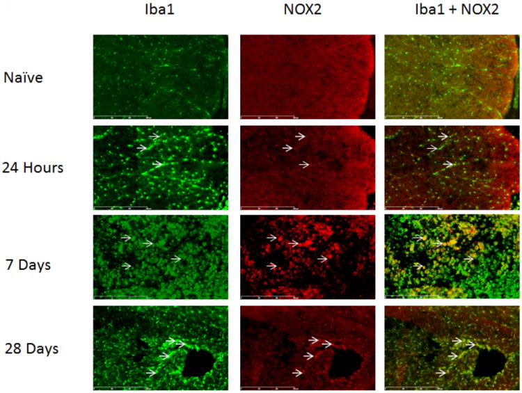
Microglia/macrophages expressed NOX2 in the spinal cord at the lesion epicenter, within regions formerly occupied by white matter. Cells labeled with the microglial marker Iba1 (green) were positive for NOX2 (red, arrows) in injured spinal cord tissue. NOX2 staining increased by 7 days-post injury, but decreased by 28 days. Bar=300μm.