
Figure 3.
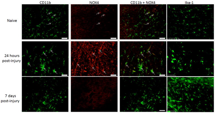
Microglia/macrophages expressed NOX4 in the spinal cord at the lesionepicenter, within regions formerly occupied by white matter. Both naïve and injured animals had CD11b positive (green) and NOX4 positive (red) cells (arrows). When merged, colabeled cells were yellow. To demonstrate consistent microglia/macrophage labeling with CD11b, adjacent sections were immunolabeled with Iba-1 and imaged at the same magnification, demonstrating similar labeling patterns. Bar= 50μm.