
Figure 4.
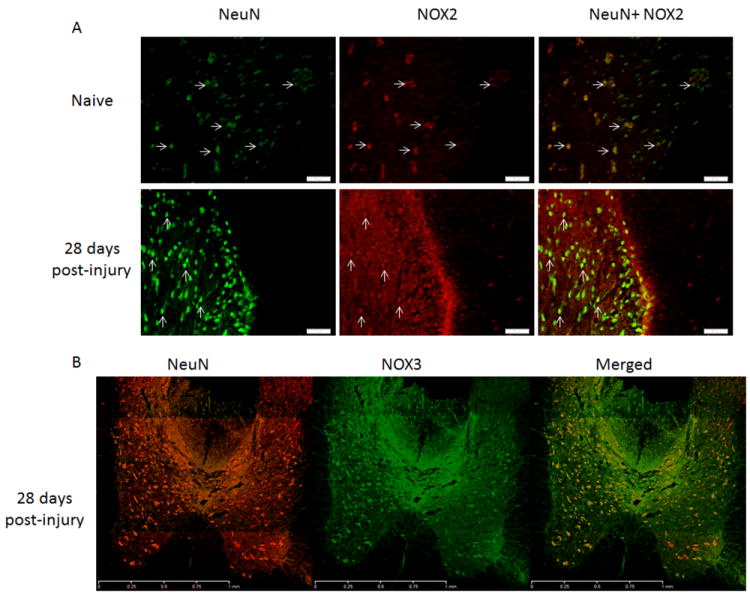
Neurons in the spinal cord expressed NOX2 and NOX3 in the dorsal horns 3mm caudalto the injury site. NOX2 (red) positive cells were also NeuN+ (green, arrows) in naïve and 28 day injured spinal cord tissue (A; bar = 50μm). NeuN positive cells (red) were found to express NOX3 (green) at all time points post-injury, with no obvious change in expression profile. Image B shows the gray matter 3mm caudal to the injury site at 28 days post-injury; bar = 1mm).