
Figure 5.
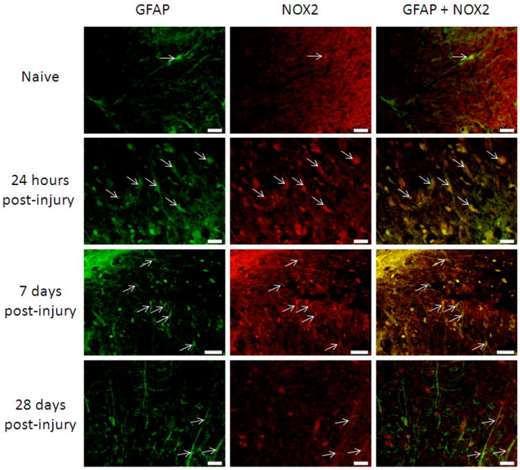
Astrocytes in the spinal cord were NOX2 positive in the spinal cord 3mm rostral to the lesion site, in white matter regions. GFAP (green) positive cells were also stained with an antibody against NOX2 (red; arrows) in naïve tissue and at 24 hours, 7 days and 28 days post-injury. Qualitatively, there appeared to be an increase in GFAP+/NOX2+ cells by 24 hours and 7 days post-injury that decreased by 28 days post-injury. Bar=20μm.