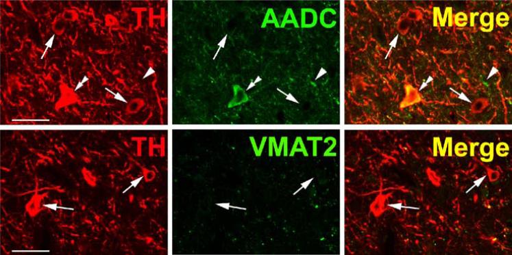
Fig. 8.
Heterogeneous phenotypes of TH + neurons in the rhesus monkey periventricular hypothalamus. Horizontal panels (rows) represent single co-stained sections analyzed by confocal microscopy. Upper panel: TH-positive neurons coexpressing AADC (double arrow heads) and TH-positive/AADC-negative neurons (arrows) are present. Note AADC-positive/TH-negative fiber (arrow head). Lower panel: TH-positive neurons lack VMAT2 (arrows). Note VMAT2-positive/TH-positive and VMAT2-positive/TH-negative puncta. Occasional immunopositivity in the region of the nucleus for the cytoplasmic antigen AADC is attributed to superimposition of cytoplasm above or below the nucleus in these sections. Bars are 30 μm.

