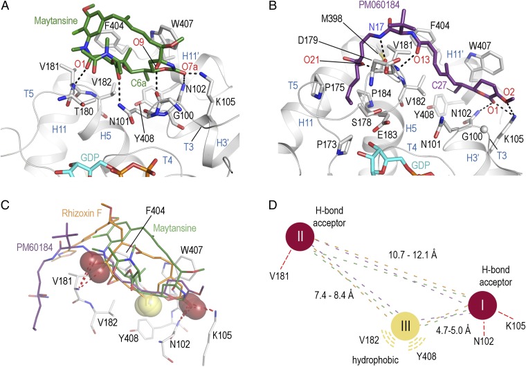
Fig. 2.
Structures of the tubulin–maytansine and tubulin–PM060184 complexes and pharmacophore model. (A) Close-up view of the tubulin–maytansine complex. Maytansine is in green stick representation. β-tubulin is displayed as gray ribbon. Key residues forming the interaction with the ligand are in stick representation and are labeled. Hydrogen bonds are highlighted as dashed black lines. (B) Detailed view of the tubulin–PM060184 complex. The ligand is displayed as violet-purple sticks. (C) Superposition of the binding sites of rhizoxin F (orange), maytansine (green), and PM060184 (violet-purple) highlighting the three common interaction points I, II, and III with β-tubulin. Hydrogen bond acceptors are highlighted as red spheres; the methyl groups forming the hydrophobic interaction are highlighted as yellow spheres. (D) Schematic drawing of the common pharmacophore for ligand binding to the maytansine site, using the same color code as in C.

