Fig. 1.
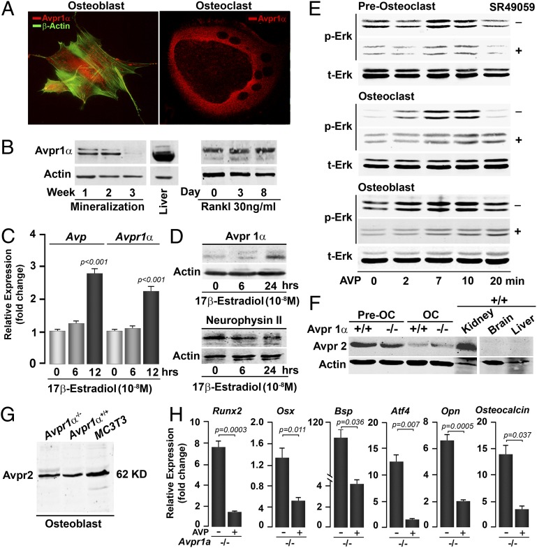
Bone cells express Avprs. Immunofluorescence micrographs (A) and Western immunoblotting (B) show the expression of Avpr1α in osteoblasts and osteoclasts, and as a function of osteoblast (mineralization) and osteoclast (with Rankl) differentiation. The expression of Avp (ligand) and Avpr1α (receptor) in osteoblasts is regulated by 17β-estradiol, as determined by quantitative PCR (C) and Western immunoblotting (D). (Magnification: A, 63×.) Because Avp is a small peptide, its precursor neurophysin II is measured. Statistics: Student t test, P values shown compared with 0 h. Stimulation of Erk phosphorylation (p-Erk) as a function of total Erk (t-Erk) by Avp (10−8 M) in osteoclast precursors (preosteoclasts), osteoclasts (OC), and osteoblasts establishes functionality of the Avpr1α in the presence or absence of the receptor inhibitor SR49059 (10−8 M) (E). Western immunoblotting showing the expression of Avpr2 in preosteoclasts, OCs (F), and osteoblasts (G) isolated from Avpr1α−/− mice, as well as in MC3T3.E1 osteoblast precursors (G). Functionality of Avpr2 was confirmed by the demonstration that cells from Avpr1α−/− mice remained responsive to AVP in reducing the expression of osteoblast differentiation genes, namely Runx2, Osx, Bsp, Atf4, Opn, and Osteocalcin (quantitative PCR, P values shown) (H). Only relevant bands from Western blots are shown, with gaps introduced where empty lanes are excised to conserve space.

