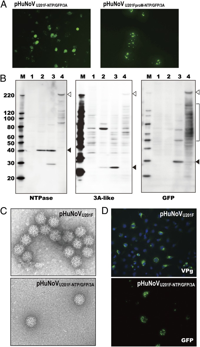
Fig. 4.
Analyses of GFP reporter constructs pHuNoVU201F-NTP/GFP/3A and pHuNoVU201FproM-NTP/GFP/3A. (A) Images of GFP expression from (Left) pHuNoVU201F-NTP/GFP/3A–transfected and (Right) pHuNoVU201FproM-NTP/GFP/3A–transfected COS7 cells at 24 hpt. (B) Analysis of proteolytic cleavage of polyprotein translated from pHuNoVU201F-NTP/GFP/3A. M represents molecular mass markers. Lane 1 shows mock-transfected cells, lane 2 shows pHuNoVU201F-transfected cells, lane 3 shows pHuNoVU201F-NTP/GFP/3A–transfected cells, and lane 4 shows pHuNoVU201FproM-NTP/GFP/3A–transfected cells. Black arrowheads show the mature protein. White arrowheads represent uncut polyprotein and brackets show intermediate cleaved precursor proteins. Antibodies used to detect the proteins are shown below the blots. (C) Progeny HuNoV virions purified from pHuNoVU201F- and pHuNoVU201F-NTP/GFP/3A–transfected cultures. (D, Upper) VPg was detected by IF in pHuNoVU201F-transfected COS7 cells that were fixed at 24 hpt and stained with the VPg mAb and Alexa Fluor 488-labeled anti-mouse IgG. Nuclei were counterstained with DAPI. (D, Lower) GFP signal was detected 24 hpt after transfection of RNA isolated from progeny virus from supernatants of pHuNoVU201F-NTP/GFP/3A–transfected cells.

