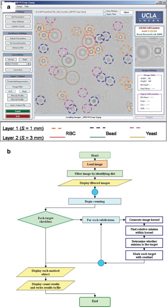
Figure 3.
a: LUCAS characterization results of a two-layered heterogeneous mixture are illustrated. Each layer of the mixture, located at S = 1.0 μm and S = 3.0 mm, contains 10 μm diameter micro-beads, red blood cells, and yeast cells (S. Pombe), all suspended in 1× PBS solution. Different colored solid and dashed circles mark the location and the height of each cell/particle type located within the sample volume—see the figure legend. b: Simplified flow-chart of the decision algorithm is shown. [Color figure can be seen in the online version of this article, available at www.interscience.wiley.com.]

