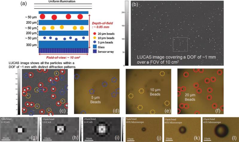
Figure 6.
a: A multi-layered object that is composed of 3 planes of micro-beads (5, 10, and 20 μm diameter) over a depth-of-field of ~0.85 mm. b: LUCAS image acquired for the object shown in (a). c: Zoomed version of the LUCAS image taken from the white frame within (b). Each micro-object exhibits a uniquely different diffraction pattern, and the solid red, dotted yellow and dashed blue circles represent 20, 10, and 5 μm diameter beads, respectively. A single LUCAS image, as shown in (b and c), shows all the particles with distinct patterns within a dept-of-field of ~1 mm and over a FOV of 10 cm2. On the other hand, a conventional microscope requires three consecutive images to monitor all these different planes of micro-objects. d–f: Demonstrate three microscope images (taken with a 10× microscope objective) that illustrate the individual planes of the multi-layered object shown in (a). Note that in each one of these microscope images, the other micro-particles are missing due to the limited depth-of-field of the microscope. g–l: The zoomed versions of the LUCAS images and the microscope images (under 40×) corresponding to 5, 10, and 20 μm diameter beads. [Color figure can be seen in the online version of this article, available at www.interscience.wiley.com.]

