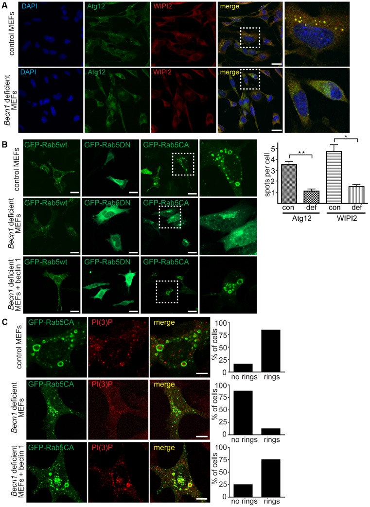
Figure 5. Autophagosome formation and endosomal expansion caused by constitutively active Rab5 and endocytosis are disrupted in Becn1 deficient MEFs.
A. Reduction of Atg12 and WIPI2 [56] puncta formation in Becn1 deficient MEFs. DAPI, endogenous Atg12, WIPI2. White dashed boxes represent zoomed regions. Scalebars = 20 µm. ‘con’ is control MEFs, ‘def’ is beclin 1 deficient MEFs. Quantification of an average of 91 cells per condition from three separate experiments (con: 91, 110, 110; def: 64, 84, 87). Bars represent mean +/− s.e.m. (n = 3); Atg12 p = 0.0039; WIPI2 p = 0.0183 using a one-tailed t-test. B. Rab5-CA mutant is mislocalized in Becn1 deficient MEFs and rescued with re-introduction of beclin 1. MEF cells were transfected with Rab5wt, CA or DN-GFP and visualized. White dashed boxes represent zoomed regions. Scale bars = 20 µm. C. PI(3)P is recruited to expanding endosome sites in control MEFs but no endosome expansion associated with Rab5CA-GFP was found in Becn1 deficient MEFs. MEF cells were transfected with Rab5CA-GFP, fixed, stained with PI(3)P antibody and visualized. Scalebars = 10 µm. Quantification of percentage of cells with no rings or rings (at least 30 transfected cells from at least 3 separate experiments). Control MEFs (7 cells with no rings, 37 cells with rings) vs. Becn1 deficient MEFs (36,5) significantly different (p<0.0001) and Becn1 deficient MEFs (36,6) vs. Becn1 revertant MEFs (11,33) significantly different (p<0.0001) using Fisher's exact test (two-sided).

