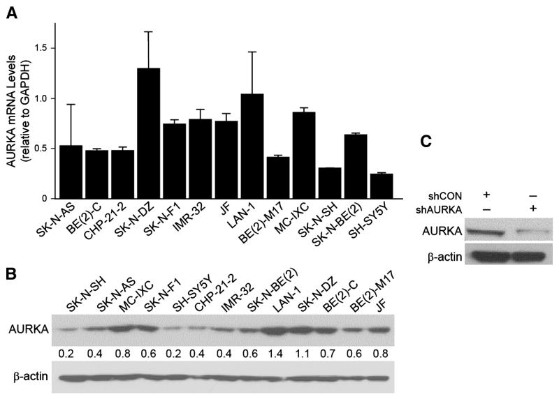
Figure 1. Constitutive mRNA and protein levels AURKA in neuroblastoma cell lines and AURKA silencing in BE(2)-C cells.
(A) Endogenous AURKA mRNA transcription activity in 13 cell lines. GAPDH was used as an internal control. (B) AURKA protein expression was examined by immunoblotting. The numbers indicate the relative expression of AURKA to β-actin measured with densitometry. (C) Immunoblotting confirmed the decrease in the protein levels after targeted silencing (shAURKA). β-actin was used as a loading control for (B) and (C).

