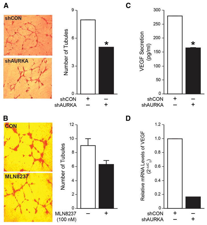
Figure 4. Inhibition of AURKA reduced angiogenesis in vitro and decreased VEGF mRNA levels and secretion.
(A) HUVECs cultured in cell culture supernatant from BE(2)-C/shAURKA cells resulted in decreased tubule formation when compared to cells grown in supernatant from BE(2)-C/shCON cells. (B) HUVECs cultured in supernatant from BE(2)-C cells treated with MLN8237 resulted in decreased tubule formation when compared to cells grown in supernatant from BE(2)-C cells treated with vehicle alone. (C) BE(2)-C/shCON and BE(2)-C/shAURKA cells were seeded in a 6-well plate, then serum-starved for 48 h. Cell culture supernatant was then collected and VEGF ELISA was performed. (D) AURKA silencing decreased mRNA levels of VEGF by ∼6 fold as assessed by real Time-PCR. GAPDH was used as an internal control. (mean ± SEM; *=p<0.05 vs. control).

