
Figure 5. Silencing AURKA decreased N-Myc expression and inhibited nuclear translocation.
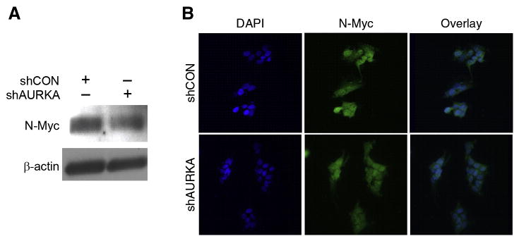
(A) N-Myc expression was decreased in BE(2)-C/shAURKA in comparison to control cells, BE(2)-C/shCON, as assessed by immunoblotting. β-actin was used as a loading control. (B) Immunofluorescence analysis demonstrated decreased nuclear translocation of N- Myc (red) in BE(2)-C/shAURKA cells when compared to controls. DAPI (blue) was used as a nuclear stain.