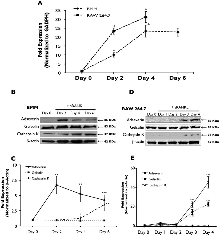
Figure 2. Adseverin expression is up-regulated during OCG in response to sRANKL.
A) Quantitative real-time PCR analysis was used to quantify Ads gene expression in osteoclast cultures derived from BMMs (black diamonds) and RAW macrophages (black circles). Results are expressed as fold expression versus Day 0 and are normalized against GADPH ± SEM. In BMM-derived osteoclasts, Ads expression was significantly up-regulated by 10-fold after 2 days and 23-fold after 4 days. No difference was noted between Day 4 and Day 6 cultures. In RAW-derived osteoclasts, Ads gene expression was significantly up-regulated by 24-fold after 2 days and 32-fold after 4 days. B) Immunoblot analysis was used to quantify protein expression in BMMs and RAW macrophage-derived osteoclast cultures. Results are expressed as fold expression versus Day 0 and normalized against ß-actin ± SD. In BMM-derived osteoclasts, Ads protein expression was significantly up-regulated by 6-fold in Day 2 cultures. 5-fold and 4-fold increases in expression were noted in Day 4 and 6 cultures, respectively. The decrease in expression in days 4 and Day 6 was not found to be statistically significant. Gelsolin expression was not altered during OCG in response to sRANKL. Cathepsin K expression was significantly increased in response to sRANKL after a 4 day stimulation with sRANKL. D) Similarly in RAW macrophage-derived osteoclast cultures, Ads was up-regulated 17-fold and 23-fold in Day 3 and Day 4 cultures respectively. Gelsolin expression was not altered during OCG, and Cathepsin K expression was significantly increased in Day 3 and 4 cultures. C & E) Quantification of immunoblots (* p<0.05, ** p<0.01, *** p<0.001, n = 3).

