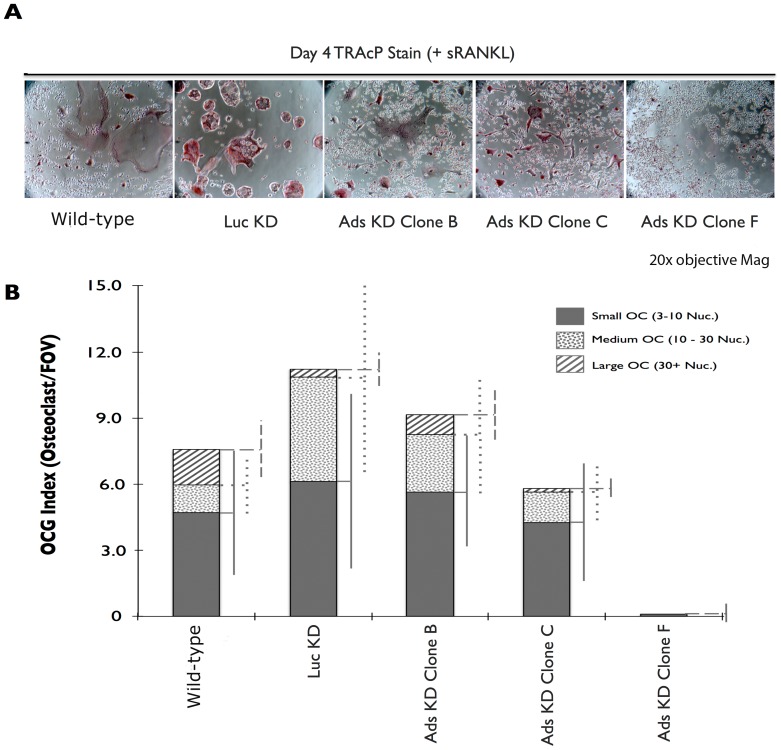
Figure 5. Adseverin knockdown Clone F displayed the greatest defect in osteoclast formation.
A) 2.5×10⁵ RAW macrophages were seeded onto 6-well tissue culture plates and stimulated for 4 days with sRANKL. Representative photomicrographs of TRAcP stained osteoclasts were taken at 20× objective magnification. A profound lack of osteoclasts was observed in Ads KD Clone F. B) The number of osteoclasts per random field of view (OCG index) was quantified. The number of osteoclasts is represented as three distinct populations of small (3–10 nuclei/osteoclast, solid grey column), medium (5–7 nuclei/osteoclast, dotted column) and large (30+ nuclei/osteoclast, dashed column) osteoclasts (n≥10). Ads KD Clones B and C experienced marginal and statically non-significant reductions in the number of large osteoclasts when compared to WT cultures. No statistically significant difference in OCG was observed between the WT and Luc KD cultures. Ads KD Clone F macrophages failed for form multinucleated osteoclasts.

