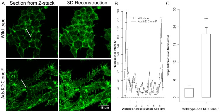
Figure 9. Adseverin knockdown alters F-actin architecture and cellular morphology in osteoclast precursors.
A) WT RAW and Ads KD osteoclasts precursors were stimulated with sRANKL for two days, fixed and stained with green- fluorescent phalloidin to localize F-actin. B) F-actin intensity was plotted against the diameter of a cell (white line, Fig 9A). Well-defined cortical actin rings were noted in the periphery of WT cells as noted by high intensity fluorescence peaks at the cell boundaries, 0.5 µm and 6 µm. Fluoresce intensity across Ads KD cells was ill defined and lacked the characteristic peaks seen in the WT cells. Ads KD had a motile phenotype characterized by dense actin rich structure and significantly increased number of filopodial extensions at the cell periphery when compared to WT. C) The number of filopodial extensions of each cell type was determined. WT cells (leftmost column) had an average of 3 (+/−1) and Ads KD Clone F cells (rightmost column) had an average of 22 (+/−2) such membrane extensions.

