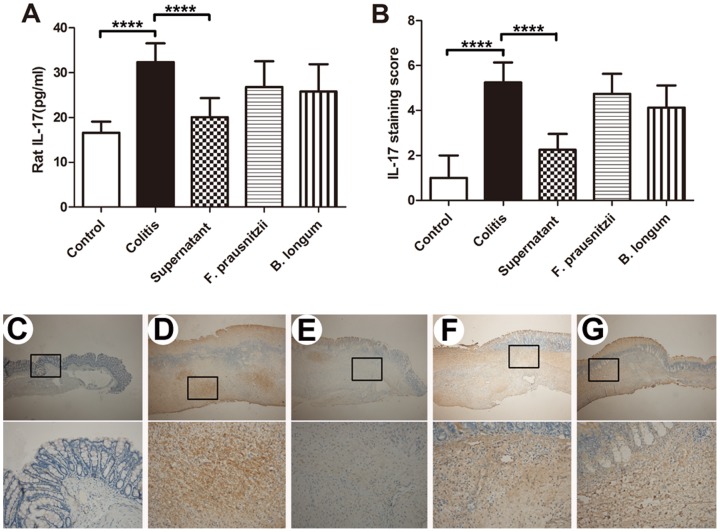
Figure 3. IL-17 protein expression in rat plasma and colon.
Plasma IL-17 concentrations in the rat (A). IL-17 protein immunohistochemical staining in rat colon (B). n = 7–8. Data are the mean ± SD. ****P <0.001. Representative immunohistochemical staining of IL-17 in rat colon mucosa in the control (C), colitis (D), supernatant (E), F. prausnitzii (F), and B. longum (G) groups. Upper and lower panel magnifications are ×40 and ×200, respectively.

