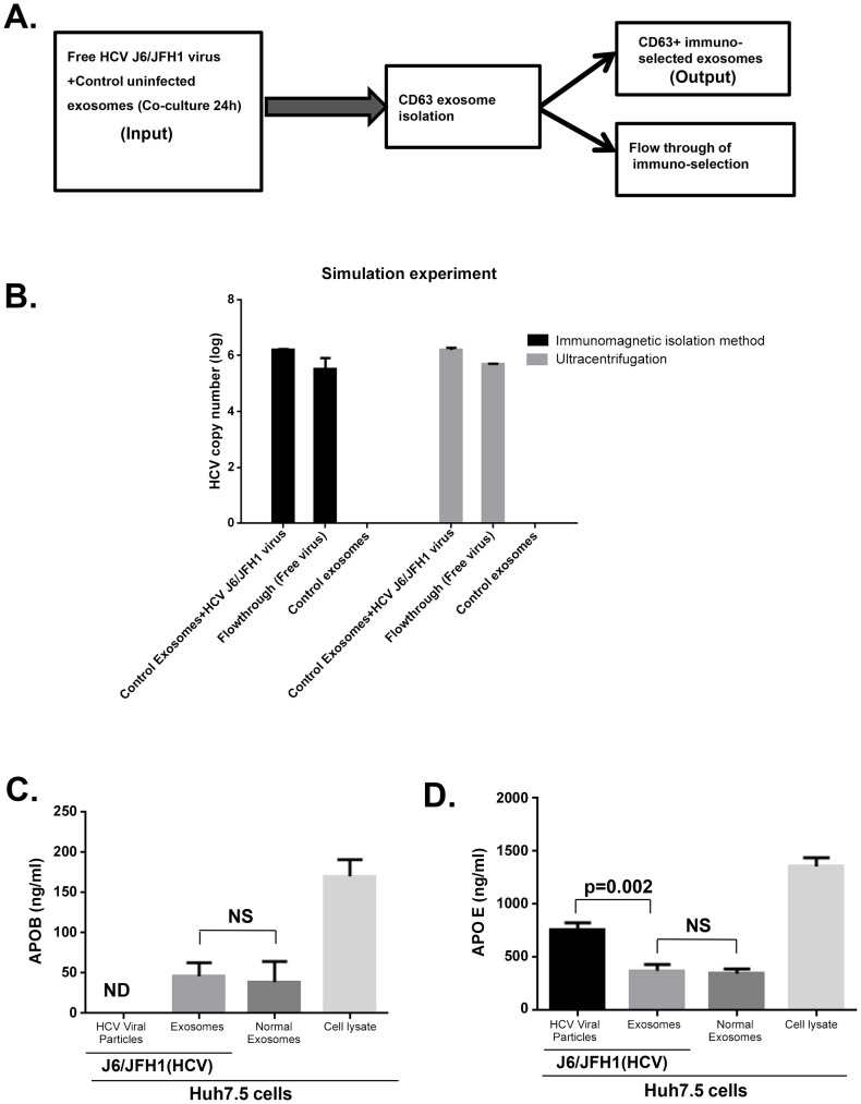
Figure 2. Simulation study ruling out viral carryover of cell free HCV virus and purity of isolated exosome.
(A) Comparative schematic flow diagram of exosome isolation by Exoquick and ultracentrifugation with additional CD63 immuno-magnetic selection. (B) Control uninfected exosomes were mixed with free HCV virus suspension and after 24 hour co-culture, samples were divided into two portions for (1) total RNA extraction or (2) immuno-magnetic CD63 isolation of the exosomes followed by total RNA extraction. Extracted RNA was analysed for HCV RNA content by quantitative real-time PCR. Results are representative of 4 indipendent experiments. (C &D) Exosomes and free HCV virus were isolated as detailed in our methods. Equal numbers of isolated exosomes and free HCV virus were then lysed in RIPA protein extraction buffer. Extracted total protein from exosomes and cell free virus as indicated was subjected to APOE and APOB ELISA analysis according to the manufacturers' protocol. Results are representative of 3 independent experiments with p<0.05 considered statistically significant usisng ANOVA analysis with GraphPad prism 5.0 software.

