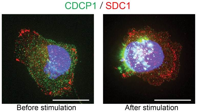
Figure 4. An adhesion molecule, Syndecan-1 (SDC1), was not associated with CDCP1 and did not respond to the stimulation of CDCP1.
HS5 cells were fixed before and after stimulation, and stained for CDCP1 (green), SDC1 (red) and nuclei (blue). SDC1 localized to the basal surface of the cells, and were especially concentrated in the focal adhesion areas in both control and stimulated cells. CDCP1 shifted toward the tip of the cells after stimulation, whereas SDC1 did not. White staining indicates CDCP1 localizing over the nucleus. Original objective, X60. Scale bars, 20 µm.

