
Figure 1.
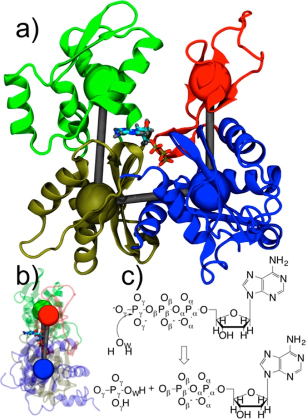
(a) Structure of G-actin with four subdomains colored differently and coarse-grained variables depicted as colored spheres. Subdomain (SD) 1 is in blue, SD2 in red, SD3 in gold, and SD4 in green. ATP and magnesium ion are also depicted. (b) Side view of actin with SD2–SD1–SD3–SD4 dihedral angle in flat, F-actin conformation. (c) ATP hydrolysis reaction with atom labels.