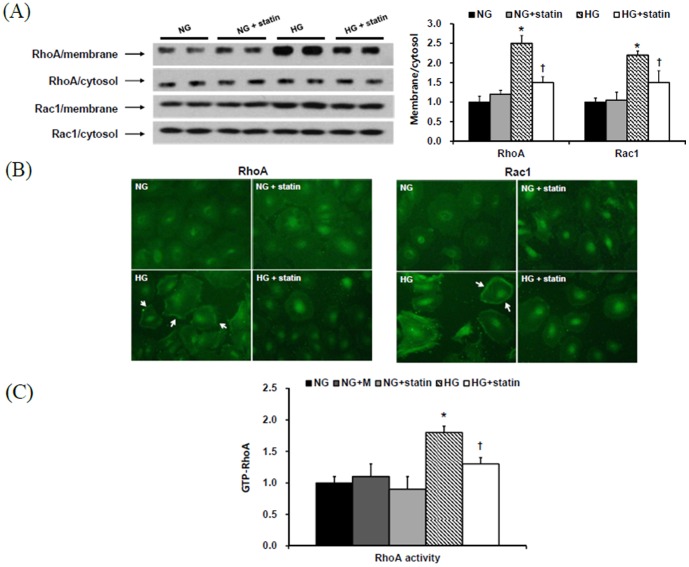
Figure 4. RhoA1 and Rac1 protein expression in the membrane and cytosol fractions of HPMCs exposed to 5.6 mM glucose (NG), NG + mannitol (94.4 mM, NG+M), NG+1 µM simvastatin (NG + statin), high glucose (100 mM, HG), or HG+1 µM simvastatin (HG + statin).
(A) The protein expression of RhoA and Rac1 were significantly increased in the membrane fraction of HG-stimulated HPMCs compared to NG cells, and simvastatin significantly attenuated the increases in RhoA and Rac1 expression in the membrane fraction of HPMCs exposed to HG. *; p<0.05 vs. NG, †; p<0.05 vs. HG. (B) An immunofluorescence study revealed that HG provoked the translocation of RhoA and Rac1 from the cytosol to the membrane fraction, and simvastatin treatment inhibited this translocation of RhoA and Rac1 induced by HG (×40). (C) The levels of Rho kinase were significantly increased in HG-treated HPMCs than in NG cells, and these changes were significantly abrogated by simvastatin. *; p<0.05 vs. NG, †; p<0.05 vs. HG.

