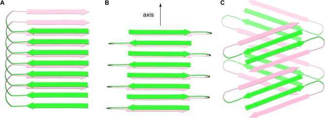
Figure 7.

Cartoon representations of fibrils formed by Aβ. (A) Parallel β-sheet fibril composed of U-shaped turns in a staggered arrangement, observed for Aβ1–40.2c (B) Antiparallel β-sheet fibril composed of U-shaped turns, observed for the Iowa mutant Aβ1–40.27 (C) Fibril-like assembly of oligomers composed of β-hairpins, that we propose from the X-ray crystallographic structure of macrocyclic β-sheet peptide 3. The green and pink colors represent the central and C-terminal regions of Aβ.
