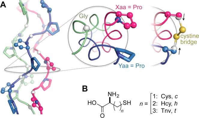
Figure 1.

(A) (PPG)10 trimer displaying Xaa (balls and sticks), Yaa (sticks), and Gly positions (white balls). Positioning of Xaa and Yaa residues is shown in a cross-section. Application of a cystine bridge here pulls Cβ atoms inward and away from their original positions (black arrows), indicating a strained linker. (B) Cysteine analogues considered in disulfide bridges in this study. All models were generated with PyMOL v1.3, unless noted otherwise.
