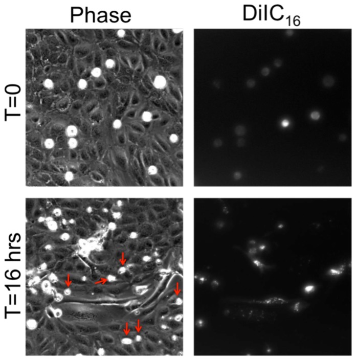
Figure 2. MDA-MB-231 incorporation causes detachment and rounding of some endothelial cells.
Phase contrast (left) and DiIC16 fluorescence (right) images of MDA-MB-231 cells plated onto an untreated HUVEC monolayer, at time points immediately after plating (top) and after 16 hours of interaction with the endothelium (bottom). Red arrows point to phase-white cells that do not emit fluorescence; these are endothelial cells that have been forced out of the monolayer and thus have detached and become rounded. Scale bar is 25 µm and applies to all images.

