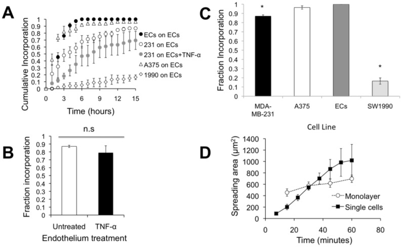
Figure 3. Incorporation of MDA-MB-231 does not depend on whether the endothelium is activated by TNF-α.
(A) Cumulative fraction of ECs or MDA-MB-231 cells (231), SW1990 (1990), and A375 cells incorporated into the endothelium as a function of time after plating. Data points represent mean ± SEM for at least 3 independent experiments (N>20 cells for each experiment). (B) Final fraction of MDA-MB-231 cells incorporated into the untreated or TNF-α-treated endothelium after 15 hours. Bars represent mean, while error bars represent SEM of at least 3 independent experiments. P>0.05 between these values indicates there is no statistical difference (n.s.). (C) Final fraction of MDA-MB-231 breast cancer cells, ECs, A375 melanoma cells, and SW1990 pancreatic cells incorporated into the endothelium after 15 hours. Bars represent mean, while error bars represent SEM of at least 3 independent experiments. (*) indicates significance (P<0.05) when compared to ECs. (D) Plot of spreading area versus time reveals differences in spreading dynamics for MDA-MB-231 cells spreading onto a fibronectin-coated coverslip (“single cells”) or into an untreated endothelium (“into monolayer”).

