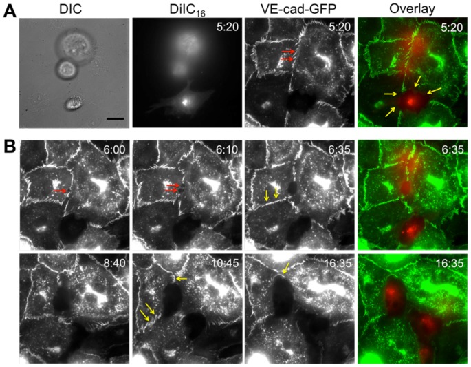
Figure 7. Cancer cell incorporation initiates by dislocating VE-cadherin at endothelial cell junctions.
DiIC16-labeled MDA-MB-231 cells were plated onto endothelial cells expressing VE-cadherin-GFP (VE-cad-GFP). (A) Shown are differential interference contrast (DIC), DiIC16 (red) fluorescence, and VE-cadherin-GFP (green) fluorescence, and overlay images. At this time point, one MDA-MB-231 has already incorporated into the endothelium (yellow arrows), and the VE-cadherin-GFP is still intact in the location directly below another MDA-MB-231 cell that has not yet begun to incorporate (red arrows). (B) Fluorescence timelapse sequence of a DiIC16-labeled MDA-MB-231 cell (red) incorporating into an endothelium expressing VE-cadherin-GFP (green). Length of time after plating MDA-MB-231 cells on the endothelium is indicated in the upper right corner of each image in hour:minute format. Scale bar in panel A (DIC image) is 10 µm and applies to all images in this figure.

